Lang:
⚕ Clinician Reference KDIGO 2024 🇵🇭 Philippines Context

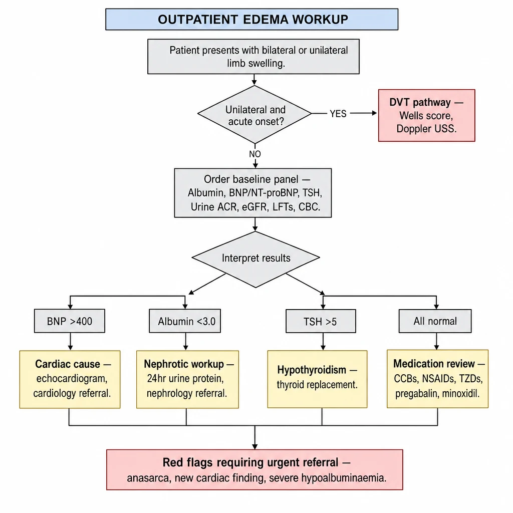
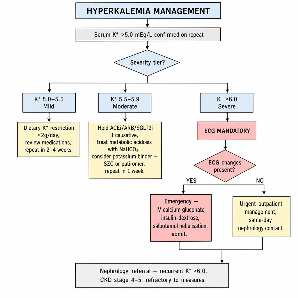
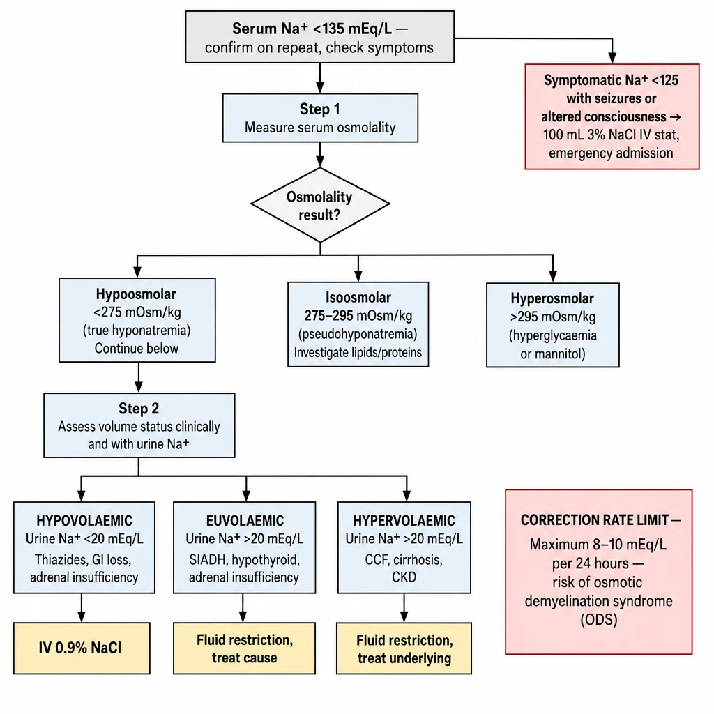
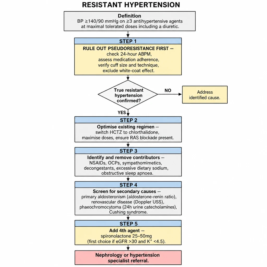
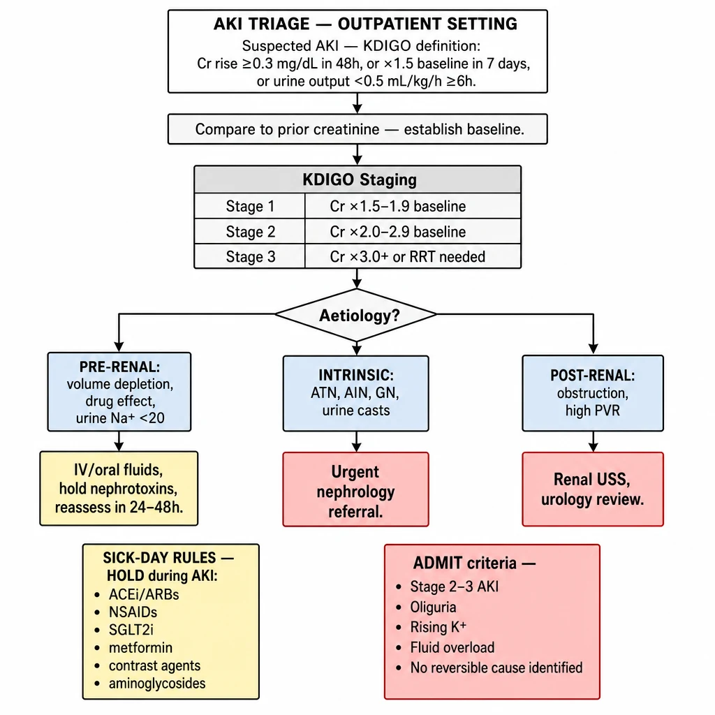
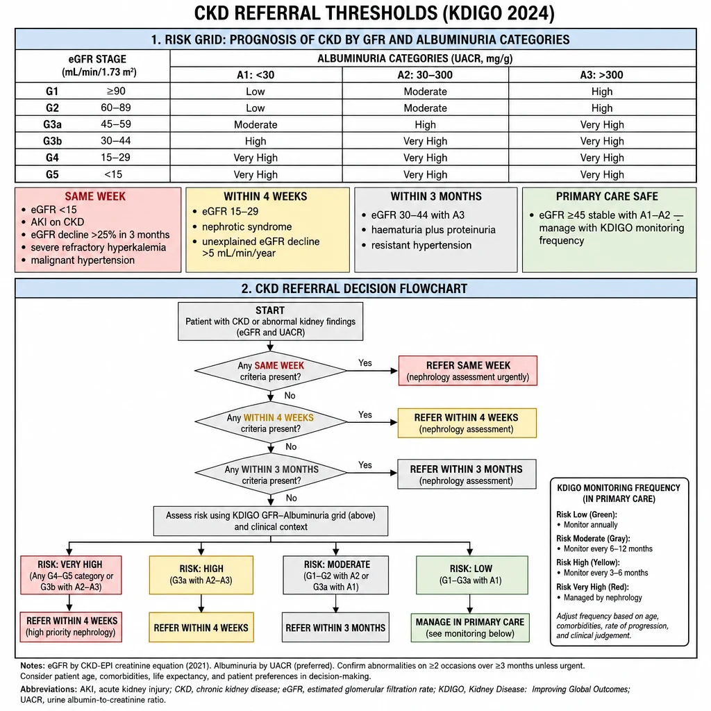
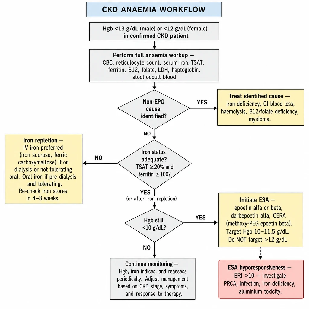
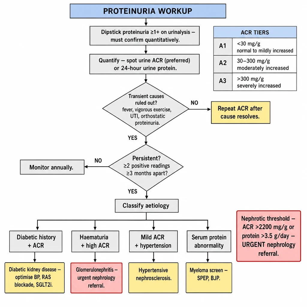
Practical Outpatient Algorithms
9 rapid decision pathways for nephrology & general medicine

Designed for GPs and IM residents managing common renal and metabolic problems in the outpatient setting. Each algorithm distills current KDIGO, ACC/AHA, and local DOH guidance into actionable decision steps.

W. G. M. Rivero, MD, FPCP, DPSN Last reviewed: May 2026 Internal Medicine · Nephrology
Clinical use only. These algorithms are decision-support tools for licensed practitioners. They do not replace clinical judgement, complete history-taking, or appropriate specialist referral. Always individualise management to the patient.
Clinical reference overview — 9 outpatient decision algorithm cards for nephrology and general medicine, KDIGO 2024 9 rapid decision algorithms at a glance — edema workup through metabolic acidosis correction. Each pathway distills KDIGO 2024, ACC/AHA, and local DOH guidance into outpatient-ready clinical steps.
1

Outpatient Edema Workup

Bilateral pitting edema is among the most common outpatient presentations. Identify the mechanism before prescribing diuretics.

Outpatient edema workup decision algorithm — unilateral DVT pathway, BNP/albumin/TSH branches, medication causes, red flags for urgent referral Algorithm 1 overview — Edema Workup. Begin with unilateral vs bilateral; order the targeted panel (BNP, albumin, TSH, ACR, eGFR); branch to cardiac, nephrotic, thyroid, or drug-induced cause. Anasarca or new cardiac finding = urgent referral.
Trigger
Patient presents with limb swelling or weight gain
First characterise: pitting vs non-pitting · unilateral vs bilateral · acute (<72 h) vs chronic · distribution (ankle only / below knee / anasarca)
Decision
Is the edema unilateral and acute?
Yes → DVT until proven otherwise

Apply Wells DVT score. If ≥2 points or high clinical suspicion → bilateral compression Doppler USS same day. If negative and score low → consider cellulitis, Baker's cyst ruptured, compartment syndrome, lymphedema (non-pitting, rubbery).

No → Bilateral / Chronic → Continue below

Bilateral edema almost always has a systemic cause. Proceed with mechanism-based workup.

Action
Order baseline panel for bilateral edema
Labs: Serum albumin · BNP or NT-proBNP · TSH · Urine ACR (spot) · LFTs (ALT, AST, bilirubin, PT) · eGFR/creatinine · CBC
Vitals review: BP, weight trend, O₂ saturation
Decision
What does the panel show?
BNP > 400 pg/mL

Cardiac cause likely. Request echo. Refer cardiology if new finding. Optimise loop diuretic (furosemide). Check electrolytes and renal function.

Albumin < 3.0 g/dL

Hypoalbuminaemia. Check urine protein (nephrotic if >3.5 g/day). Check LFTs for cirrhosis. Check total protein + SPEP if myeloma suspected. → Refer nephrology if nephrotic range.

TSH > 5 mIU/L

Hypothyroid edema. Classic non-pitting myxedema. Start levothyroxine; edema resolves with euthyroid state over weeks.

Decision
All labs normal — is the patient on any of these medications?
High-yield medication causes: Amlodipine / CCBs NSAIDs / mefenamic acid Pioglitazone / TZDs Pregabalin / gabapentin Minoxidil Corticosteroids Hormones / OCP
Yes → Medication-induced

Withdraw or switch offending agent. CCB-induced edema: switch to ARB + thiazide combination. Monitor for resolution over 2–4 weeks.

No → Venous insufficiency

Most common in chronic bilateral ankle edema without systemic cause. Confirm with venous duplex. Manage: compression stockings, leg elevation, sodium restriction.

Admit If
Red flags requiring urgent escalation
Anasarca with albumin <2.0 g/dL · BNP >1,000 (acute decompensated HF) · O₂ saturation <94% at rest · Creatinine acutely rising · BP >180/120 with edema · New ascites with altered sensorium (hepatic encephalopathy)

Quick Reference — edema Mechanism Map

  • Increased hydrostatic pressure: CHF, venous insufficiency, hypoalbuminaemia
  • Decreased oncotic pressure: nephrotic syndrome, cirrhosis, malnutrition
  • Increased capillary permeability: inflammation, allergy, infection
  • Lymphatic obstruction: lymphedema (non-pitting), filariasis
  • Sodium/water retention: CKD, medications, SIADH
  • Tip: Amlodipine is the most missed cause in the Philippines — always check CCB use first
2

Hyperkalemia Management

Outpatient-focused. Any K⁺ ≥ 6.0 mEq/L requires an ECG before a management decision. Type IV RTA (hyporeninaemic hypoaldosteronism) is the most common cause in CKD/DKD outpatients.

Hyperkalemia management algorithm — severity tiers 5.0–5.5 / 5.5–5.9 / ≥6.0, ECG mandatory above 6.0, dietary restriction, potassium binders, nephrology referral criteria Algorithm 2 overview — Hyperkalemia Management. Classify by severity: Mild 5.0–5.5, Moderate 5.5–5.9, Severe ≥6.0 with mandatory ECG. Outpatient steps: dietary K⁺ restriction, hold offending agents, treat acidosis, consider patiromer or SZC. K⁺ ≥6.0 + ECG changes = emergency.
Trigger
Serum K⁺ above 5.0 mEq/L on routine labs
Rule out pseudohyperkalaemia first: haemolysed sample · prolonged transport (>2 h) · extreme leukocytosis or thrombocytosis (WBC >100,000 or plt >1,000,000) · fist clenching during draw. Repeat if clinical picture doesn't match.
Decision
What is the confirmed K⁺ level?
LevelSeverityImmediate Step
5.0–5.4 mEq/LMildDietary review + medication audit. Recheck in 2–4 weeks.
5.5–6.0 mEq/LModerateECG. Dietary restriction. Consider potassium binder. Reduce/hold RAASi if appropriate.
> 6.0 mEq/LSevereECG immediately. If any ECG changes → ADMIT. If ECG normal and patient asymptomatic → may manage outpatient with close follow-up.
Admit If
Any ECG changes present → emergency admission
Peaked symmetrical T waves (early) · PR prolongation · Wide QRS (>120 ms) · Sine-wave pattern or VF (pre-arrest)
In-hospital: IV calcium gluconate (membrane stabilisation) → insulin + dextrose → sodium bicarbonate (if acidosis) → kayexalate/SPS enema → dialysis if refractory.
Action
Outpatient management steps (mild–moderate, no ECG changes)
1. Dietary restriction
  • Target <40 mEq/day (≈ 1,500 mg K⁺)
  • Avoid: saging (banana), abokado (avocado), kamote, mongo, coconut water
  • Leaching technique for vegetables (peel, cut, boil in large water, discard water)
2. Medication review
  • ACEi / ARB reduce dose or switch if K⁺ >5.5 with CKD
  • NSAIDs stop — reduce aldosterone activity
  • TMP-SMX stop — blocks ENaC like amiloride
  • K-sparing diuretics hold spironolactone if K⁺ >5.5
  • Loop diuretics increase dose if volume overloaded
Action
Potassium binders — when and which
Use if K⁺ persistently >5.5 mEq/L despite dietary restriction and medication optimisation, especially when RAASi continuation is clinically important (CKD progression, heart failure).

Sodium zirconium cyclosilicate (SZC / Lokelma): 10 g TID × 48 h (acute), then 5–10 g QD maintenance. Onset 1 h. Preferred in Philippines (available). Raises serum HCO₃ slightly — beneficial in CKD acidosis.
Patiromer (Veltassa): 8.4 g QD with food. Onset 7–12 h. Not interchangeable with SZC. Use away from other medications (chelates). Limited availability in PH.
Sodium polystyrene sulphonate (Kayexalate): older agent; avoid in ileus risk; PO or enema. Still widely used in PH due to cost.
Action
Treat contributing metabolic acidosis
Metabolic acidosis shifts K⁺ extracellularly (↑ K⁺ by ~0.6 mEq/L per 0.1 unit ↓ in pH). Correcting HCO₃ to ≥22 mEq/L with oral sodium bicarbonate (see Algorithm 9) reduces K⁺ by 0.3–0.6 mEq/L in CKD patients — often sufficient for mild hyperkalaemia.
Refer
Nephrology referral criteria for hyperkalaemia
K⁺ persistently >5.5 despite maximal outpatient therapy · Recurrent admissions for severe hyperkalaemia · K⁺ >5.5 with eGFR <30 (needs CKD co-management) · Suspected type IV RTA requiring diagnostic workup (renin, aldosterone levels)

Quick Reference — Hyperkalaemia Memory Aids

  • C-BIG-K⁺ Drop (acute treatment order): Calcium gluconate → Bicarbonate → Insulin+Glucose → Kayexalate/SZC → Dialysis
  • Type IV RTA: low renin + low aldosterone; K⁺ 5.0–6.0; mild NAGMA; eGFR 30–60; DKD most common cause
  • RAAS inhibitors: necessary for cardio-renal protection — do not stop without binder trial first
  • Recheck K⁺ within 1–2 weeks after any intervention
  • If on SZC: recheck K⁺ in 48–72 h during loading phase
  • Avoid raw/unprocessed bananas, kamote leaves, and coconut water in dialysis patients
3

Hyponatremia Management

Serum Na⁺ <135 mEq/L. Classify by osmolality first, then volume status. The correction rate is as important as the target — overcorrection causes osmotic demyelination syndrome (ODS).

Hyponatremia management 3-step algorithm — serum osmolality classification, volume status branching, correction rate limits ≤10 mEq/24h, symptomatic Na+ below 125 emergency protocol Algorithm 3 overview — Hyponatremia Management. Step 1: serum osmolality (hypoosmolar / isoosmolar / hyperosmolar). Step 2: volume status (hypovolaemic → SIADH → hypervolaemic). Step 3: correction rate — max 8–10 mEq/L per 24h to prevent ODS. Symptomatic Na⁺ <125 = 100 mL 3% NaCl bolus.
Trigger
Serum Na⁺ <135 mEq/L found on labs
Assess symptoms: asymptomatic vs mild (nausea, headache) vs moderate (vomiting, confusion) vs severe (seizures, coma, respiratory arrest). Symptomatic hyponatraemia → hospital.
Decision
Step 1: Measure serum osmolality
Osm >290 mOsm/kg (Hypertonic)

Translocational hyponatraemia. Hyperglycaemia most common. Correct Na⁺ by +1.6 mEq/L per 100 mg/dL glucose above 100. Treat glucose — Na⁺ normalises.

Osm 280–290 mOsm/kg (Isotonic)

Pseudohyponatraemia. Extreme hyperlipidaemia or hyperproteinaemia. Measure Na⁺ on blood gas analyser (direct ISE). No treatment of Na⁺ needed.

Osm <280 mOsm/kg (Hypotonic)

True hyponatraemia. Proceed to Step 2: assess volume status.

Decision
Step 2: Assess volume status (clinical + urine studies)
Hypovolaemic (dry)
  • Signs: ↓ skin turgor, orthostasis, ↑ BUN/Cr ratio, dry mucosa
  • Urine Na <20: extra-renal loss (vomiting, diarrhoea, burns, third-spacing)
  • Urine Na >20: renal loss (diuretics, salt-wasting, Addison's)
  • Treatment: Isotonic saline 0.9%. Correct volume deficit first — Na⁺ will follow.
Euvolaemic (normal volume)
  • No edema, normal BP, mildly ↓ urine output
  • SIADH most common cause
  • Always rule out: hypothyroidism (TSH), cortisol deficiency (morning cortisol), medications
  • Medications causing SIADH: SSRIs, SNRIs, carbamazepine, TMP-SMX, PPIs, amiodarone, cyclophosphamide, oxytocin
  • Treatment: Fluid restriction 800–1,000 mL/day. Salt tabs. If refractory: tolvaptan (V2-receptor antagonist) — specialist-initiated only.
Hypervolaemic (edematous)
  • edema + ascites present
  • Causes: CHF, cirrhosis, nephrotic syndrome
  • Urine Na typically <20 (effective arterial volume depletion despite edema)
  • Treatment: Fluid restriction + treat underlying cause. Loop diuretic if CHF. Do not give saline.
Warning
Correction rate limits — the most important rule
Chronic hyponatraemia (>48 h or unknown duration): Maximum correction = 8–10 mEq/L per 24 hours · 18 mEq/L per 48 hours.
Faster correction → Osmotic demyelination syndrome (ODS): central pontine myelinolysis. Dysarthria, dysphagia, quadriparesis, locked-in syndrome. Irreversible.
High-risk for ODS: Alcohol use disorder · malnutrition · hypokalaemia · baseline Na⁺ <120 mEq/L · liver disease.
Acute hyponatraemia (<48 h): Can correct at 1–2 mEq/L/h initially until symptoms resolve (usually +5 mEq/L sufficient for seizures/coma).
Admit If
Symptomatic or severe hyponatraemia
Seizures, coma, obtundation, vomiting with aspiration risk → ADMIT immediately.
Emergency: 3% NaCl 150 mL IV over 20 min → repeat ×2 if no improvement → target +5 mEq/L in first hour → reassess. Stop 3% NaCl once symptomatic improvement or Na⁺ reaches 130 mEq/L.

SIADH Diagnostic Criteria (all must be met)

  • Serum osmolality <280 mOsm/kg · Urine osmolality >100 mOsm/kg (inappropriately concentrated)
  • Urine Na⁺ >40 mEq/L (renal Na wasting despite hyponatraemia) · Clinically euvolaemic
  • Normal thyroid function · Normal adrenal function · Not on diuretics
  • Common causes: CNS disease, pulmonary disease (TB, pneumonia, lung cancer), malignancy (SCLC), drugs
Na⁺ Correction Rate Estimator
4

Resistant Hypertension

BP ≥ 140/90 mmHg on ≥ 3 maximally tolerated antihypertensive agents from different classes, including a diuretic. Pseudoresistance is far more common than true resistant hypertension — rule it out first.

Resistant hypertension 5-step clinical algorithm — pseudoresistance exclusion with 24h ABPM, regimen optimisation, secondary cause screening including primary aldosteronism, spironolactone as 4th agent Algorithm 4 overview — Resistant Hypertension. Step 1: rule out pseudoresistance (white-coat, adherence, cuff error — check 24h ABPM). Steps 2–4: optimise regimen, screen drug/lifestyle contributors, evaluate for secondary causes. Step 5: add spironolactone 25–50 mg as preferred 4th agent.
Trigger
Clinic BP ≥ 140/90 on ≥ 3 antihypertensives at adequate doses
Includes patients on ≥ 4 drugs with controlled BP (controlled resistant HTN). This accounts for ~10–20% of treated hypertensives.
Decision
Step 1: Rule out pseudoresistance — the most important step
Measurement errors
  • Wrong cuff size (most common) — use large cuff for arm circumference >33 cm
  • Talking, crossing legs, unsupported arm during measurement
  • Measure after 5 min rest in seated position
  • Check both arms — use higher reading arm
White-coat / Non-adherence
  • White-coat HTN: Home BP monitoring (HBPM) target <135/85 mmHg at home. ABPM gold standard (daytime avg <135/85).
  • Non-adherence: Pill count, pharmacy records, directly observed dosing, urine drug screen for antihypertensives (thiazide metabolite).
  • Suboptimal doses or inappropriate drug combinations (two ARBs, two CCBs)
Action
Step 2: Optimise current regimen before adding agents
Essential questions:
(1) Is a diuretic included? Thiazide (HCTZ/chlorthalidone) for eGFR ≥30; loop diuretic (furosemide BID) if eGFR <30 or fluid overload.
(2) Are drugs at maximum tolerated doses?
(3) Are doses timed correctly? Evening dosing of one agent improves nocturnal dipping.
(4) Single-pill combinations improve adherence — preferred where available in PH.
Action
Step 3: Identify and eliminate lifestyle and drug contributors
Lifestyle factors
  • Sodium intake >3 g/day (Filipino diet averages 5–6 g) — dietary recall critical
  • Alcohol >14 units/week raises BP significantly
  • OSA — very prevalent in PH; partner-reported snoring, BMI >30, Epworth ≥10 → sleep study
  • Weight gain, sedentary lifestyle
Pressor medications
  • NSAIDs (mefenamic acid, ibuprofen) — very common in PH OTC use
  • Decongestants (pseudoephedrine, phenylephrine)
  • OCP (ethinylestradiol-containing pills)
  • Energy drinks (caffeine + taurine)
  • Corticosteroids (chronic oral prednisone)
  • Calcineurin inhibitors (cyclosporine, tacrolimus)
Decision
Step 4: Screen for secondary hypertension
CausePrevalenceClueScreen
Primary hyperaldosteronismMost common (~20%)K⁺ low or low-normal; adrenal incidentaloma; severe or early-onset HTNAldosterone-to-renin ratio (ARR) — off interfering drugs ×2 weeks. ARR >30 with Aldo >15 → positive. Refer endocrine.
Obstructive sleep apnoea~30–40%Snoring, witnessed apnoeas, daytime sleepiness, obesity, thick neckEpworth Sleepiness Scale; overnight oximetry; polysomnography. CPAP reduces SBP ~5–10 mmHg.
Renal artery stenosis~3–5%Flash pulmonary edema; worsening creatinine with RAASi; abdominal bruit; atherosclerotic risk; FMD (young women)Renal Doppler USS. CTA/MRA if Doppler inconclusive. Refer vascular/nephrology.
Phaeochromocytoma0.1–0.6%Paroxysmal headache + sweating + palpitations triad; episodic hypertension; weight loss24-h urine metanephrines + catecholamines (preferred) or plasma metanephrines. CT abdomen if positive.
Cushing's syndromeRareTruncal obesity, proximal myopathy, purple striae, moon face, buffalo hump, easy bruisingOvernight 1 mg dexamethasone suppression test. Cortisol >1.8 μg/dL → positive. Refer endocrine.
Action
Step 5: Add 4th-line agent (true resistant HTN confirmed)
First choice: Spironolactone 25–50 mg daily (PATHWAY-2 trial: most effective 4th agent; reduces SBP ~8–9 mmHg vs placebo). Check K⁺ and eGFR at 4 weeks. Avoid if K⁺ >5.0 or eGFR <30.
If spironolactone not tolerated (gynaecomastia, hyperkalaemia):
Eplerenone 25–50 mg (more selective, less anti-androgenic) · Doxazosin 4–8 mg (α-blocker) · Bisoprolol 5–10 mg if not on BB · Moxonidine (central α-agonist, but inferior evidence).
Avoid clonidine (rebound hypertension on missed doses — dangerous in outpatient use).
Refer
When to refer to nephrology or hypertension specialist
BP uncontrolled after 4-drug regimen + lifestyle + adherence confirmed · Confirmed secondary cause (aldosteronism, RAS) · eGFR <45 with resistant HTN (likely CKD-driven; needs nephrology co-management) · Consider renal denervation evaluation at tertiary centres

4-Drug Sequence for Resistant HTN (after pseudoresistance excluded)

  • Step 1: RAS blocker (ACEi or ARB) + CCB (amlodipine) + Thiazide (chlorthalidone preferred over HCTZ) — maximise all doses
  • Step 2: Add loop diuretic if eGFR <30 or edema present (switch thiazide to furosemide BID)
  • Step 3: Add spironolactone 25 mg → titrate to 50 mg (PATHWAY-2 evidence)
  • Step 4: Add doxazosin or bisoprolol based on patient profile · Refer if still uncontrolled
5

AKI Triage — Outpatient Setting

Acute Kidney Injury (KDIGO): rise in serum creatinine ≥0.3 mg/dL within 48 h, or ×1.5 baseline within 7 days, or urine output <0.5 mL/kg/h for ≥6 h. Always compare to a prior creatinine — do not assume baseline from a single elevated value.

AKI triage outpatient algorithm — KDIGO staging table, pre/intrinsic/post-renal aetiology columns, sick-day rules ACEi/ARBs/NSAIDs/SGLT2i, admit criteria Stage 2-3 AKI oliguria rising potassium Algorithm 5 overview — AKI Triage. KDIGO staging: Stage 1 (×1.5–1.9), Stage 2 (×2.0–2.9), Stage 3 (×3.0+). Aetiologies: pre-renal (urine Na⁺ <20), intrinsic (casts, AIN, GN), post-renal (USS). Sick-day rules: hold ACEi/ARBs, NSAIDs, SGLT2i. Admit: Stage 2–3, oliguria, rising K⁺, no reversible cause.
Trigger
Acutely elevated creatinine or oliguria on outpatient visit
First: retrieve previous creatinine. Check eHR, prior labs, old results from other facilities. A creatinine of 2.0 mg/dL may be AKI (if baseline 0.8) or chronic CKD (if stable for 2 years). This distinction drives everything.
Decision
Classify AKI severity (KDIGO staging)
StageCreatinine criterionUrine outputTypical approach
Stage 1×1.5–1.9 baseline or ↑ ≥0.3 mg/dL<0.5 mL/kg/h × 6–12 hOutpatient if pre-renal likely; remove offending agents; 48 h recheck
Stage 2×2.0–2.9 baseline<0.5 mL/kg/h × 12 hStrong consideration for admission; close monitoring at minimum
Stage 3×3.0 or Cr >4.0 mg/dL or initiation of KRT<0.3 mL/kg/h × 24 h or anuria × 12 hAdmit — nephrology consult urgent
Decision
Classify aetiology: Pre-renal / Intrinsic / Post-renal
Pre-renal (most common outpatient)
  • History: vomiting, diarrhoea, poor oral intake, bleeding, diuretic use, heat exposure
  • Exam: decreased turgor, orthostasis (↑ HR or ↓ SBP ≥20 on standing), dry mucosa
  • Labs: BUN/Cr >20 · FENa <1% (unreliable if on diuretics — use FEUrea <35% instead) · Urine SG >1.020
  • Treatment: Oral/IV rehydration. Stop NSAIDs, diuretics. Hold ACEi/ARB temporarily.
Intrinsic renal
  • ATN: Recent contrast, aminoglycosides (amikacin), prolonged pre-renal, rhabdomyolysis (CPK elevation)
  • GN: Active urine sediment (dysmorphic RBCs, RBC casts), proteinuria, hematuria, hypertension, systemic symptoms (rash, arthralgias, haemoptysis)
  • AIN: Recent drug (NSAIDs, PPIs — especially high-dose, beta-lactam antibiotics) · rash + eosinophilia + fever (Triad present in only 10%) · FENa >1%
Post-renal (obstructive)
  • Classic: BPH (most common in Filipino men >60), bladder neck obstruction, pelvic malignancy, bilateral ureteric stones, neurogenic bladder
  • Exam: palpable bladder, suprapubic fullness, flank pain
  • Test: Bedside bladder USS (post-void residual >300 mL) or renal USS (hydronephrosis bilateral). Relief of obstruction often restores function rapidly.
Action
Sick-day rules — medications to hold during AKI
NSAIDs — reduce renal blood flow via prostaglandin inhibition; worst in volume depletion
ACEi / ARB — hold if volume depleted, K⁺ >5.5, or creatinine rising >25% within 2 weeks of initiation
Metformin — hold if Cr >1.5 mg/dL in women, >1.7 in men, or any AKI (lactic acidosis risk)
SGLT2 inhibitors — hold during any acute illness / hospitalisation (euglycaemic DKA risk)
Contrast agents — schedule IV contrast procedures only when renal function stable
Aminoglycosides — adjust dose per eGFR; avoid in AKI unless no alternative
Beta-blockers — continue; do not stop abruptly
Statins — continue
Admit If
Indications for hospital admission from the outpatient clinic
AKI Stage 2 or 3 · Oliguria or anuria · K⁺ >6.0 mEq/L or any ECG change · Volume overload with respiratory compromise · Creatinine rising >0.5 mg/dL in 24 h · Unknown aetiology requiring workup (GN, vasculitis) · Inability to take oral medications or maintain hydration at home · Social circumstances precluding safe outpatient monitoring

Outpatient AKI Monitoring Plan (Stage 1 Pre-renal — safe for outpatient)

  • Remove offending agents (NSAIDs, diuretics, RAASi temporarily)
  • Oral rehydration: 2–2.5 L oral fluids per day if not contraindicated by cardiac status
  • Recheck creatinine, electrolytes, and urine output assessment in 48–72 h
  • Clear return precautions: ↓ urine output, worsening edema, confusion, chest pain, BP >180/120
  • If not improving or worsening at 48-h recheck → escalate to admission
FeNa / FeUrea Calculator — Pre-renal vs Intrinsic AKI
Creatinine:
Urea nitrogen:

On diuretics? Add BUN values to calculate FeUrea (more reliable):

6

CKD Referral Thresholds

Based on KDIGO 2024 CKD guidelines. Referral is based on eGFR, albuminuria, rate of decline, and complications — not eGFR alone. Many patients with eGFR 45–60 can be safely managed in primary care with shared-care guidance.

CKD referral thresholds infographic — KDIGO 2024 heat-map grid eGFR G1-G5 by albuminuria A1-A3, four urgency tiers: urgent same-week, within 4 weeks, within 3 months, primary-care safe Algorithm 6 overview — CKD Referral Thresholds (KDIGO 2024). The G×A heat-map assigns green / yellow / amber / red risk. Urgency tiers: Same week (eGFR <15, AKI-on-CKD, malignant HTN) → 4 weeks (eGFR 15–29, nephrotic) → 3 months (eGFR 30–44 + A3) → Primary care safe (eGFR ≥45 stable, A1–A2).
Trigger
CKD confirmed: eGFR <60 mL/min/1.73m² or urine ACR ≥30 mg/g on ≥2 occasions, ≥3 months apart
Confirm CKD staging using both eGFR (G1–G5) and albuminuria (A1–A3) — the G×A heatmap guides risk and referral urgency. A single creatinine is insufficient; trends over time matter most.
Urgent (<1 week)
Refer urgently — same week or emergency department
eGFR <15 mL/min/1.73m² (G5) — KRT planning required urgently
Suspected rapidly progressive GN: hematuria + proteinuria + creatinine rising over weeks (RPGN may progress to dialysis-dependence within weeks if untreated)
Hyperkalaemia >6.5 mEq/L not responding to emergency measures
Hypertensive emergency with new renal impairment
Pulmonary edema with new oliguria
Suspected hemolytic uraemic syndrome (microangiopathic hemolytic anemia + thrombocytopaenia + AKI)
Prompt (<1 month)
Refer within 4 weeks
eGFR <30 mL/min/1.73m² (G4) — KRT preparation, AVF planning, anemia management, metabolic acidosis
eGFR decline >5 mL/min/1.73m²/year (rapid progressor) or >10 mL/min in 5 years
Persistent urine ACR >300 mg/g despite optimised ACEi/ARB + BP control
Recurrent AKI episodes (>3 per year)
Difficult-to-manage HTN with CKD (suspected renovascular or secondary cause)
Inherited kidney disease: PKD, Alport syndrome, FSGS in young patient
Unexplained proteinuria >1 g/g without diabetes or HTN
Routine (<3 months)
Refer within 3 months or co-manage with nephrology guidance
eGFR 30–44 (G3b) with any of: anemia Hgb <11, metabolic acidosis HCO₃ <22, CKD-MBD (↑PTH, ↑PO₄, ↓Ca), persistent hyperkalaemia
Diabetic nephropathy G3a+ with ACR >300 despite maximal dual RAS blockade
CKD with suspected glomerular disease (hematuria + proteinuria, abnormal sediment)
CKD requiring biopsy for diagnosis (proteinuria without clear cause)
Monitor
Safe to manage in primary care — monitor per KDIGO frequency
eGFR ≥45 (G1–G3a) + ACR <30 (A1) + controlled BP + controlled diabetes → annual nephrology review may be deferred
Review frequency by G×A risk category: Low (G1–G2, A1) = annual; Moderate (G3a, A1) = annual; High (G3a A2, G3b A1) = 2×/year; Very High = 3–4×/year
Monitor: BP, eGFR, ACR, electrolytes (K⁺, HCO₃), Hgb, Ca, PO₄, PTH (if eGFR <45)

Rule of thumb: Refer when you would question your next step

  • eGFR <30 → always refer (G4/G5)
  • ACR >300 persistent → always refer
  • Rapidly declining eGFR → always refer
  • Any doubt about aetiology → refer for biopsy consideration
  • AVF planning: Refer vascular surgery when eGFR <25 and HD is likely trajectory — earlier creation means better maturation
  • Pre-dialysis education: Start when eGFR <30 — modality choice, diet, access, transplant evaluation
  • Transplant referral: eGFR <20 and declining — pre-emptive transplant is the best option
CKD-EPI 2021 eGFR Calculator
Creatinine units:
7

CKD anemia Workflow

anemia of CKD is a diagnosis of exclusion — always rule out iron deficiency, hemolysis, B12/folate deficiency, and GI blood loss before attributing anemia to EPO deficiency. The sequence: replete iron → reassess → consider ESA.

CKD anemia workflow algorithm — full workup excluding other causes, iron status TSAT ferritin thresholds, IV iron preferred, ESA sequence epoetin darbepoetin CERA, Hgb target 10-11.5, ESA hyporesponsiveness ERI above 10 Algorithm 7 overview — CKD anemia Workflow. Workup first: CBC, iron studies, reticulocytes, B12/folate, LDH, stool OB. Rule out non-EPO causes. Iron repletion (IV preferred): TSAT <20% or ferritin <100. If Hgb still <10 after iron → ESA (epoetin, darbepoetin, CERA). Target Hgb 10–11.5 g/dL. ERI >10 = investigate hyporesponsiveness.
Trigger
Hgb <13 g/dL (males) or <12 g/dL (females) in a patient with CKD
CKD anemia typically normochromic normocytic. Develop earlier in CKD-DM. Nearly universal in G4–G5. Begin investigation when Hgb falls below threshold — do not wait for symptoms.
Action
Step 1: Full anemia workup — before any treatment
CBC + differential + reticulocyte count · Serum iron, TIBC, ferritin, TSAT
Vitamin B12 · Folate · LDH + haptoglobin + peripheral smear (hemolysis screen)
Faecal occult blood test (GI blood loss) · Stool H. pylori (common cause of iron deficiency in PH)
If hemolysis suspected: Direct Coombs, G6PD deficiency screen
Decision
Step 2: Is iron deficiency present?
PopulationIron deficient ifTreatment
CKD non-dialysisFerritin <100 ng/mL or TSAT <20%Oral iron first-line
CKD on HDFerritin <200 ng/mL or TSAT <20%IV iron preferred (poor oral absorption, inflammation)
CKD on PDFerritin <100 ng/mL or TSAT <20%Oral or IV iron
Oral iron options: Ferrous sulfate 325 mg TID between meals (elemental iron 65 mg/tab) · Ferrous fumarate · Carbonyl iron (better tolerated GI-wise)
IV iron options (available in PH): Iron sucrose (Venofer) 200 mg over 30 min × 5 doses · Ferric carboxymaltose (Ferinject) 500–1000 mg single infusion (off-label in CKD; widely used)
Recheck iron stores at 3 months. A Hgb response of ≥1 g/dL in 4–6 weeks confirms iron-responsive anemia.
Decision
Step 3: After iron repletion — is Hgb still <10 g/dL?
Yes → Consider ESA
  • Target Hgb: 10–11.5 g/dL (KDIGO 2024 — do NOT target >13 g/dL: ↑CVD risk, stroke)
  • ESA options (PH): EPO-alfa (Eprex) 50–100 IU/kg SC 3×/week · Darbepoetin-alfa 0.45 mcg/kg SC Q2W · EPO-beta (NeoRecormon)
  • Check reticulocyte response at 2–4 weeks. Titrate dose by 25–50% if Hgb not rising.
  • Do not use ESA if active malignancy (promotes tumour growth), uncontrolled HTN, or pure red cell aplasia
No → Monitor

Iron-responsive anemia corrected. Recheck Hgb every 3 months. Maintain TSAT >20% and ferritin >100. Continue oral iron maintenance if oral route tolerated.

Warning
ESA hyporesponsiveness — investigate before escalating dose
Defined as: No Hgb rise ≥1 g/dL despite EPO dose >300 IU/kg/week (or darbepoetin equivalent).
Common causes: Iron deficiency (most common — recheck stores) · Active infection or inflammation (hepcidin ↑) · Hyperparathyroidism (PTH >500 suppresses erythropoiesis) · Folate/B12 deficiency · Aluminium toxicity (rare) · Dialysis inadequacy (Kt/V <1.2) · Pure red cell aplasia (anti-EPO antibodies — check if on IV EPO >3 months with sudden resistance)
Transfuse
Blood transfusion — avoid if possible, especially in transplant candidates
Avoid transfusion in pre-transplant patients — HLA sensitisation reduces transplant eligibility dramatically.
Consider transfusion if: Hgb <7 g/dL with symptoms (dyspnoea at rest, angina, altered sensorium) · Hgb <8 g/dL peri-operatively · Acute hemorrhage requiring immediate correction.
If transfusion required in transplant candidate → document, inform transplant team, request leuco-depleted filtered blood.

CKD anemia Targets — KDIGO 2024

  • Target Hgb: 10.0–11.5 g/dL for most CKD patients on ESA
  • Do NOT target Hgb >13 g/dL — TREAT (normal Hgb trial): ↑ stroke, thromboembolic events, death
  • Iron target: TSAT ≥20% + Ferritin >100 ng/mL (non-dialysis) or >200 (HD)
  • Do not give IV iron if ferritin >500 (functional iron overload risk) without reassessment
  • HIF-PHI (roxadustat, daprodustat): approved in some countries; monitor CVD risk; check local availability in PH
IV Iron Dose Calculator — Ganzoni Formula
hemoglobin units:
KDIGO 2024 target: 10–11.5 g/dL
8

Proteinuria Workup

Proteinuria on dipstick must be confirmed quantitatively and be persistent (≥2 readings ≥3 months apart) to diagnose CKD. The degree and pattern guide the diagnostic pathway.

Proteinuria workup algorithm — quantify with spot ACR A1/A2/A3 tiers, transient vs persistent definition, aetiology branches DKD hypertensive nephrosclerosis GN myeloma, nephrotic threshold ACR above 2200 urgent referral Algorithm 8 overview — Proteinuria Workup. Quantify: spot urine ACR (A1 <30 / A2 30–300 / A3 >300 mg/g). Rule out transient causes (fever, exercise, UTI, orthostatic). Persistent = ≥2 readings over 3 months. Aetiology: DKD, hypertensive nephrosclerosis, GN, myeloma (BJP). Nephrotic threshold ACR >2200 mg/g = urgent nephrology referral.
Trigger
Positive dipstick protein (≥1+) on urinalysis
Dipstick limitations: Detects only albumin (misses light chains, tubular proteins) · False positive: concentrated urine, alkaline urine, hematuria, contamination.
Confirm with: Spot urine albumin-to-creatinine ratio (ACR) — preferred, most practical in PH outpatient setting. Alternatively, 24-hour urine protein collection (gold standard but cumbersome).
Action
Quantify proteinuria (spot urine ACR)
ACR mg/g (or mg/mmol)CategoryClinical Significance
<30 mg/gA1 — Normal / mildly increasedMonitor annually if CKD risk factors present
30–300 mg/gA2 — Moderately increasedMicroalbuminuria — CKD marker; maximise ACEi/ARB + SGLT2i
>300 mg/gA3 — Severely increasedMacroalbuminuria — refer nephrology; high progression and CVD risk
>2,200 mg/gNephrotic rangeEvaluate for nephrotic syndrome — urgent nephrology referral
Decision
Is the proteinuria transient or persistent?
Repeat ACR in 2–4 weeks. Instruct patient to avoid: vigorous exercise (within 24 h), fever, UTI, menstruation, prolonged upright posture.
Orthostatic proteinuria: Common in adolescents and young adults. Protein present only in upright urine; absent in first morning void (supine overnight). Benign; confirm with split urine collection.
Transient or orthostatic

Reassure. Repeat ACR in 6–12 months. No treatment needed. If ACR normalises and remains normal — not CKD.

Persistent (≥2 occasions)

Classify aetiology and determine CKD stage with eGFR. Proceed with workup below.

Decision
What is the likely aetiology?
Diabetic nephropathy (DN)
  • Diabetes + ACR 30–300 (microalbuminuria) + NO hematuria + eGFR may be normal or ↓
  • Typically gradual onset; bilateral kidneys normal size or enlarged
  • Management: ACEi or ARB (first-line) · SGLT2i (canagliflozin, empagliflozin) · BP <130/80 · HbA1c <7% · Finerenone if ACR >300 + eGFR 25–75
  • Refer if ACR >300 or eGFR declining despite optimised therapy
Non-diabetic proteinuria
  • Check: ANA, anti-dsDNA (lupus) · ANCA (vasculitis) · C3/C4 (complement-mediated) · Anti-PLA2R (MN) · SPEP/UPEP (myeloma if >40 y/o) · Hepatitis B/C serology · HIV
  • Active urine sediment (RBC casts, dysmorphic RBCs) → GN → urgent nephrology
  • Heavy proteinuria + normal urine sediment → membranous nephropathy or FSGS → refer
Nephrotic Syndrome
Nephrotic syndrome diagnosis — always refer to nephrology
Classic triad: Proteinuria >3.5 g/day + Serum albumin <3.0 g/dL + Pitting edema (± hyperlipidaemia, lipiduria).
Causes in Filipino adults: Membranous nephropathy (most common adult-onset) · FSGS · Minimal change disease (often steroid-responsive) · Amyloidosis (if myeloma, chronic infection, or FMF) · Diabetic nephropathy (if heavy).
Complications to manage while awaiting nephrology: Thromboembolism (PE risk when albumin <2.0 — consider anticoagulation) · Infection risk (encapsulated organisms — vaccinate) · Hyperlipidaemia (statin).

Proteinuria Workup Summary — Outpatient Approach

  • Confirm: Repeat spot ACR × 2 readings, ≥3 months apart
  • Quantify: ACR (spot) or 24-h urine protein (if ACR inconclusive)
  • Characterise: Urine microscopy (casts, dysmorphic RBCs?)
  • Baseline: eGFR, electrolytes, albumin, LFTs, CBC, BP
  • Diabetic: ACEi/ARB + SGLT2i + BP control → refer if ACR >300 or declining
  • Non-diabetic + hematuria + casts → GN → urgent nephrology
  • Nephrotic range → always refer → biopsy usually required
  • ACR 30–300 stable + diabetes/HTN controlled → manage in primary care
9

Metabolic Acidosis Correction

Step-wise approach: confirm the primary disorder, calculate the anion gap (with albumin correction), classify, then treat the cause. CKD metabolic acidosis (Type IV RTA and uraemic acidosis) are the most common outpatient presentations.

Metabolic acidosis workup algorithm — confirm pH HCO3, anion gap calculation with albumin correction, high AG MUDPILES mnemonic, normal AG HARDDUP RTA types I II IV, CKD treatment oral sodium bicarbonate, pH below 7.2 referral Algorithm 9 overview — Metabolic Acidosis. Confirm: pH <7.35, HCO₃ <22. Calculate AG (Na − Cl − HCO₃; correct for albumin). High AG (MUDPILES): uremia, DKA, lactic acidosis, toxins. Normal AG (HARDDUP): hyperchloraemic — RTA types I/II/IV, diarrhoea. CKD treatment: oral NaHCO₃, target HCO₃ >22. Refer if pH <7.2.
Trigger
ABG or venous blood gas showing pH <7.35 with HCO₃ <22 mEq/L
Step 1: Confirm primary disorder and assess compensation.
Winter's formula (expected pCO₂ for pure metabolic acidosis):
Expected pCO₂ = (1.5 × HCO₃) + 8 ± 2 If measured pCO₂ < expected → concurrent respiratory alkalosis If measured pCO₂ > expected → concurrent respiratory acidosis (e.g., COPD)
Action
Step 2: Calculate anion gap (AG) and correct for albumin
AG = Na⁺ − (Cl⁻ + HCO₃⁻) Normal: 8–12 mEq/L Albumin-corrected AG = measured AG + [2.5 × (4.0 − albumin g/dL)] ↑ by 2.5 mEq/L for each 1 g/dL drop in albumin below 4.0 Delta-delta ratio (if high AG present): δδ = (AG − 12) / (24 − HCO₃) <0.4 → pure NAGMA (no AG component) 0.4–2 → pure HAGMA >2 → HAGMA + concurrent metabolic alkalosis
Decision
Step 3: Classify by anion gap
High AG metabolic acidosis (HAGMA)

MUDPILES mnemonic:

  • Methanol poisoning
  • Uraemia (CKD G4–G5)
  • DKA / alcoholic / starvation ketoacidosis
  • Propylene glycol (IV medications)
  • Isoniazid / Iron toxicity
  • Lactic acidosis (Type A: hypoperfusion; Type B: metformin, liver disease)
  • Ethylene glycol (antifreeze)
  • Salicylate toxicity

Calculate osmolar gap if toxic alcohol suspected: OG = measured Osm − calculated Osm. OG >10 → toxic alcohol.

Normal AG (NAGMA / hyperchloraemic)

Calculate urine anion gap (UAG):

UAG = UNa + UK − UCl
  • Negative UAG (−): Appropriate NH₄⁺ excretion. Extra-renal cause: diarrhoea (most common in PH), ileostomy, fistula. → Treat with oral NaHCO₃ or K-citrate.
  • Positive UAG (+): Impaired NH₄⁺ excretion → Renal tubular acidosis (RTA).
Action
RTA classification (positive UAG = impaired renal acidification)
TypeUrine pHK⁺HCO₃CauseAction
Type I (Distal)>5.5 (can't acidify)LowVariable (often <15)Sjögren's, autoimmune, amphotericin, nephrocalcinosis/stonesNaHCO₃ or K-citrate. Refer nephrology.
Type II (Proximal)<5.5 (can acidify, but leaks HCO₃)Low<15 mEq/LFanconi syndrome, myeloma, tenofovir, heavy metalsHigh-dose NaHCO₃ (often 10+ mEq/kg/day). Refer nephrology.
Type IV (Hypoaldosteronism)<5.5HighMild reduction (18–22)Most common in CKD/DKD, ACEi/ARB use — hyporeninaemic hypoaldosteronismTreat hyperkalaemia (Algorithm 2). Fludrocortisone if confirmed Addison's. Often improves with NaHCO₃ + K⁺ management.
Action
CKD metabolic acidosis — outpatient treatment (most common scenario)
Target: serum HCO₃ ≥22 mEq/L (KDIGO 2024 — slows CKD progression, preserves muscle mass, improves bone health, reduces hospitalisation).

Sodium bicarbonate (NaHCO₃):
Starting dose: 0.5–1.0 mEq/kg/day divided into 2–3 doses with meals
Tablet: NaHCO₃ 650 mg = 7.7 mEq HCO₃ (available in PH pharmacies)
Powder: Baking soda (sodium bicarbonate) 1 level teaspoon = 48 mEq — practical and inexpensive for PH patients
Titrate every 4 weeks until HCO₃ reaches 22–26 mEq/L

Caution with sodium load: Each gram NaHCO₃ = 12 mEq Na⁺ — monitor BP and edema. If fluid overload is a concern, consider potassium citrate (if K⁺ is normal or low) as a sodium-free alternative.

Benefits beyond acidosis correction: Improved muscle protein retention · Reduced phosphate release from bone · Better response to EPO · Reduced hyperkalaemia (see Algorithm 2)
Refer
When to refer metabolic acidosis to nephrology or toxicology
Severe acidaemia: pH <7.20 or HCO₃ <10 → hospital (may need IV NaHCO₃ or haemodialysis) · Lactic acidosis type A (sepsis, shock) → ICU · Toxic alcohol ingestion (methanol, ethylene glycol) → emergency department + toxicology · Type I or II RTA with nephrocalcinosis/stones or Fanconi syndrome → nephrology · CKD acidosis not responding to oral NaHCO₃ at 1 mEq/kg/day → nephrology co-management

Quick Reference — Metabolic Acidosis Formulas

  • Anion gap: Na − (Cl + HCO₃) · Normal 8–12 mEq/L
  • Albumin correction: AG + 2.5 × (4 − Alb)
  • Winter's formula: pCO₂ = (1.5 × HCO₃) + 8 ± 2
  • Delta-delta: (AG−12) / (24−HCO₃)
  • Urine AG: UNa + UK − UCl · Negative = GI loss · Positive = RTA
  • NaHCO₃ 650 mg tab = 7.7 mEq HCO₃
  • Baking soda 1 tsp = ~48 mEq NaHCO₃ (practical for home use)
  • Type IV RTA = most common CKD acidosis; hyperkalemia + mild NAGMA in DKD
Anion Gap, Delta-Delta & Bicarbonate Deficit Calculator
Albumin units:
For corrected AG. Normal = 4.0 g/dL.
Tap outside or press Esc to close